Diagram Of Animal Cell As Seen Under Electron Microscope : Illustrate only a plant cell as seen under electron ... / Transmission electron microscopy (tem) is a microscopy technique in which a beam of electrons is transmitted through a specimen to form an image.
byDelora Kirkland-
0
Diagram Of Animal Cell As Seen Under Electron Microscope : Illustrate only a plant cell as seen under electron ... / Transmission electron microscopy (tem) is a microscopy technique in which a beam of electrons is transmitted through a specimen to form an image.. There are also more intriguing shapes such as curved, spherical, concave and rectangular. The specimen is most often an ultrathin section less than 100 nm thick or a suspension on a grid. Most of the cells are microscopic in size and can only be seen under the microscope. Transmission electron microscopy (tem) is a microscopy technique in which a beam of electrons is transmitted through a specimen to form an image.
Most of the cells are microscopic in size and can only be seen under the microscope. Transmission electron microscopy (tem) is a microscopy technique in which a beam of electrons is transmitted through a specimen to form an image. There are also more intriguing shapes such as curved, spherical, concave and rectangular. The specimen is most often an ultrathin section less than 100 nm thick or a suspension on a grid.
cells under electron microscope - Google Search | Animal ... from i.pinimg.com Transmission electron microscopy (tem) is a microscopy technique in which a beam of electrons is transmitted through a specimen to form an image. Most of the cells are microscopic in size and can only be seen under the microscope. The specimen is most often an ultrathin section less than 100 nm thick or a suspension on a grid. There are also more intriguing shapes such as curved, spherical, concave and rectangular.
Most of the cells are microscopic in size and can only be seen under the microscope.
There are also more intriguing shapes such as curved, spherical, concave and rectangular. The specimen is most often an ultrathin section less than 100 nm thick or a suspension on a grid. Most of the cells are microscopic in size and can only be seen under the microscope. Transmission electron microscopy (tem) is a microscopy technique in which a beam of electrons is transmitted through a specimen to form an image.
Transmission electron microscopy (tem) is a microscopy technique in which a beam of electrons is transmitted through a specimen to form an image. Most of the cells are microscopic in size and can only be seen under the microscope. There are also more intriguing shapes such as curved, spherical, concave and rectangular. The specimen is most often an ultrathin section less than 100 nm thick or a suspension on a grid.
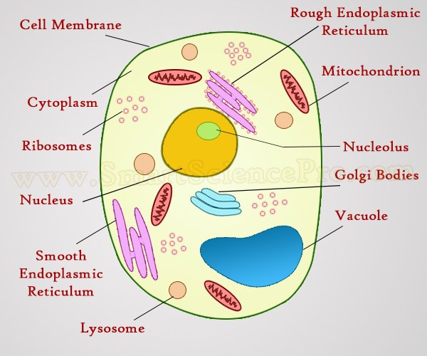
Structure of Animal Cell and Plant Cell Under Microscope ... from www.smartsciencepro.com There are also more intriguing shapes such as curved, spherical, concave and rectangular. Most of the cells are microscopic in size and can only be seen under the microscope. The specimen is most often an ultrathin section less than 100 nm thick or a suspension on a grid. Transmission electron microscopy (tem) is a microscopy technique in which a beam of electrons is transmitted through a specimen to form an image.
The specimen is most often an ultrathin section less than 100 nm thick or a suspension on a grid.
Most of the cells are microscopic in size and can only be seen under the microscope. Transmission electron microscopy (tem) is a microscopy technique in which a beam of electrons is transmitted through a specimen to form an image. The specimen is most often an ultrathin section less than 100 nm thick or a suspension on a grid. There are also more intriguing shapes such as curved, spherical, concave and rectangular.
Most of the cells are microscopic in size and can only be seen under the microscope. The specimen is most often an ultrathin section less than 100 nm thick or a suspension on a grid. There are also more intriguing shapes such as curved, spherical, concave and rectangular. Transmission electron microscopy (tem) is a microscopy technique in which a beam of electrons is transmitted through a specimen to form an image.
Illustrate only a plant cell as seen under electron ... from www.studyrankersonline.com Most of the cells are microscopic in size and can only be seen under the microscope. The specimen is most often an ultrathin section less than 100 nm thick or a suspension on a grid. Transmission electron microscopy (tem) is a microscopy technique in which a beam of electrons is transmitted through a specimen to form an image. There are also more intriguing shapes such as curved, spherical, concave and rectangular.
The specimen is most often an ultrathin section less than 100 nm thick or a suspension on a grid.
The specimen is most often an ultrathin section less than 100 nm thick or a suspension on a grid. There are also more intriguing shapes such as curved, spherical, concave and rectangular. Most of the cells are microscopic in size and can only be seen under the microscope. Transmission electron microscopy (tem) is a microscopy technique in which a beam of electrons is transmitted through a specimen to form an image.